
Immunohistochemistry double staining (Double Immunohistochemistry, Double IHC) is a pathological detection technology upgraded from conventional single immunohistochemistry staining. The core principle is to simultaneously and specifically label and localize two different target antigens on the same tissue section (paraffin section/frozen section), distinguishing signals through two chromogenic systems with distinct contrast. This can intuitively demonstrate the expression location, expression level, and co-localization relationship of two proteins. The biggest difference from fluorescence methods is that image merging is not required; the color differences between two proteins can be observed with the naked eye, and the results can be permanently preserved without concerns about signal degradation.
The underlying logic of immunohistochemistry double staining is based on antigen-antibody specific binding and enzymatic chromogenic reactions. The core challenge is to avoid cross-interference between the two staining systems and ensure that both signals are independent, specific, and free of non-specific staining. The specific principle is divided into two steps:
1. Specific recognition: Using two primary antibodies from different species origins with non-interfering specificities to respectively bind two different target antigens in the tissue section, achieving precise targeted localization of antigens.
2. Differential chromogenic detection: Paired with corresponding enzyme-labeled secondary antibodies to catalyze chromogenic reactions of different chromogen substrates, generating two insoluble pigment precipitates with distinct color contrast. Common combinations include DAB brown + Fast Red red, or cyan, golden yellow, magenta, and other chromogenic combinations can be selected according to requirements to clearly distinguish signals of two target proteins.
The basic operational framework, preliminary pretreatment, and basic chromogenic system of immunohistochemistry double staining are highly similar to traditional single immunohistochemistry tissue staining, and the sample processing logic, basic reagents, and chromogenic principles of conventional single staining can be followed. The overall process follows the basic workflow of section deparaffinization, antigen retrieval, antibody incubation, enzymatic chromogenic reaction, counterstaining and mounting. Conventional chromogenic reagents such as DAB and Fast Red are also commonly used with traditional single staining.
1. Conventional Sample Pretreatment
Deparaffinize paraffin sections to water, perform antigen retrieval through high-temperature high-pressure or microwave methods to fully expose antigen sites; inactivate endogenous peroxidase in tissues, block non-specific binding sites with blocking solution to reduce background non-specific staining, laying the foundation for subsequent dual-system staining.
2. First Round Antigen Staining and Chromogenic Detection
Apply the first primary antibody for incubation, add HRP-labeled secondary antibody for incubation after thorough washing; apply DAB substrate for chromogenic reaction, terminate the reaction with tap water when positive signals are clear. At this point, the first round target protein presents stable brown signals.
3. Double Staining Exclusive: Complete Inactivation of First Round System
This is the core operation distinguishing it from traditional single staining. After the first round of staining, a large amount of active HRP enzyme remains on the section surface. If the second round of staining proceeds directly, residual enzyme will catalyze substrate non-specific chromogenic reaction, causing whole-field false positives and background contamination. Complete quenching of residual enzyme activity is required through high-temperature boiling or high-concentration hydrogen peroxide blocking to completely isolate cross-interference between the two staining systems.
4. Double Staining Exclusive: Second Round Differential Paired Staining and Chromogenic Detection
Different from the fixed single staining system of single staining, double staining requires strict pairing with a second system without cross-reactivity: use primary antibodies from different species than the first round, paired with enzyme-labeled secondary antibodies, apply Fast Red substrate for chromogenic reaction to form red positive signals. The entire process precisely controls incubation and chromogenic duration to balance dual-color signal intensity, avoiding the first round dark signals masking weakly expressed secondary signals, or insufficient chromogenic development of secondary signals leading to target loss.
5. Sample Post-processing and Interpretation
Use hematoxylin for mild counterstaining of cell nuclei to avoid masking dual-color positive signals; after gradient dehydration, clearing, and mounting with neutral balsam, observe under microscope to distinguish single-positive and double-positive signals, complete photography and result interpretation. Compared with single staining, double staining post-processing requires consideration of the integrity of both color signals, with higher requirements for operational precision.
1. Precise in situ co-localization: Observing two protein expressions in the same cell and same tissue region can directly determine protein co-expression, adjacent expression, and differential expression relationships, completely solving the problem of multi-section alignment deviation.
2. High sample utilization rate: Only a single section is needed to complete dual-indicator detection, greatly saving precious clinical pathological samples, especially suitable for puncture samples and micro-biopsy samples.
3. More correlated results: Simultaneously obtain expression abundance and distribution location of two proteins, precisely analyze functional relationships of protein synergy and antagonism, assisting mechanism research and classification diagnosis.
4. Compatible with conventional equipment: No special fluorescence equipment required, observable with ordinary optical microscopes; chromogenic products have strong stability, and sections can be preserved for long periods and repeatedly examined.
Whether breast ductal carcinoma in situ has microinvasion is a common difficulty in pathological diagnosis. Conventional morphological observation and single-indicator immunohistochemistry staining are prone to missed diagnosis and suspicious misdiagnosis of microinvasion foci due to section deviation and field misalignment. Immunohistochemistry double staining can simultaneously label myoepithelial cells and epithelial cell markers on the same section. The commonly used optimal combination of calponin/p63 + E-cadherin clearly demonstrates the integrity of the myoepithelial layer, precisely identifying microinvasive changes of cancer cells breaking through the basement membrane. Clinical data confirm that this double staining protocol can significantly improve the diagnostic rate of breast microinvasive carcinoma, reduce the suspicious diagnosis rate, effectively solve the diagnostic challenge of difficult-to-define microinvasive lesions, and significantly improve the diagnostic accuracy of early invasive breast lesions.
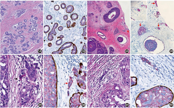
Figure 1. Normal breast tissue. Figure A shows HE staining; Figure B shows cocktail calponin/p63+E-cadherin double immunohistochemical staining
Figure 2. Breast ductal carcinoma in situ with multifocal invasion (arrows show invasion foci). Figure A shows HE staining; Figure B shows cocktail calponin/p63+E-cadherin double immunohistochemical staining
Figure 3. Breast ductal carcinoma in situ with microinvasion (arrows show microinvasion foci). Figure A shows HE staining; Figure B shows cocktail calponin/p63+E-cadherin double immunohistochemical staining
Figure 4. Breast high-grade ductal carcinoma in situ with microinvasion. Figure A shows HE staining; Figure B shows cocktail calponin/p63+E-cadherin double immunohistochemical staining
Lymphoma subtypes are complex with high morphological overlap. Relying solely on HE morphology or separate immunohistochemistry and EBER in situ hybridization makes it difficult to precisely determine the origin of positive cells, prone to ambiguous classification. Using immunohistochemistry combined with in situ hybridization double staining technology, dual determination at gene level (EBER) and protein level (CD3/CD20) can be simultaneously achieved on the same cell. Based on whether EBER-positive cells co-express T-cell marker CD3 or B-cell marker CD20, T- and B-cell-derived lymphomas can be rapidly distinguished. This double staining mode avoids tissue alignment errors of multi-section detection. Combined with the standardized staining sequence of in situ hybridization first followed by immunohistochemistry, it maximally preserves nucleic acid and protein signal specificity, providing reliable in situ molecular evidence for precise lymphoma classification.
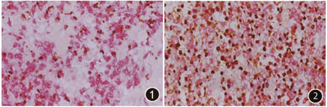
Figure 1. Experimental group NK/T cell lymphoma, EBER-positive cell nuclei show brown color, in situ hybridization; CD3-positive cell cytoplasm shows red color, EnVision method
Figure 2. Experimental group EB virus-positive diffuse large B-cell lymphoma, EBER-positive cell nuclei show brown color, in situ hybridization; CD20-positive cell membranes show red color, EnVision method
Perineural invasion of colonic adenocarcinoma is an important indicator for assessing tumor invasiveness and prognosis. However, traditional HE combined with single-indicator immunohistochemistry staining requires multi-section comparison, with low recognizability of small nerve invasion foci, time-consuming examination, and prone to missed diagnosis. Using the S-100/CKpan double staining system can specifically label nerve fibers (S-100, brown) and colonic adenocarcinoma tumor cells (CKpan, red) respectively within the same section. The dual-color contrast is clear with precise localization, intuitively demonstrating tumor cell infiltration, encirclement, and invasion range of nerves. The diagnostic results of this double staining method show good consistency with traditional methods. While ensuring diagnostic accuracy, it can significantly improve the detection capability of small and occult nerve invasion foci, while greatly shortening pathological examination time and improving overall diagnostic efficiency, suitable for routine precise assessment of nerve invasion in colonic adenocarcinoma.
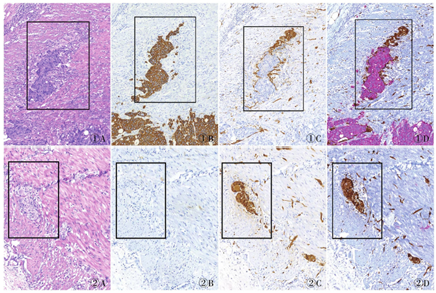
Figure 1. Colonic adenocarcinoma with visible perineural invasion, boxed area shows peripheral nerve infiltrated by adenocarcinoma. A. HE staining results; B. CKpan immunohistochemistry staining results; C. S-100 immunohistochemistry staining results; D. S-100/CKpan immunohistochemistry double staining results
Figure 2. Colonic adenocarcinoma without perineural invasion, boxed area shows peripheral nerve tissue, no adenocarcinoma infiltration. A. HE staining results; B. CKpan immunohistochemistry staining results; C. S-100 immunohistochemistry staining results; D. S-100/CKpan immunohistochemistry double staining results
The double immunohistochemistry detection system developed by EnkiLife can simultaneously detect two different antigens on the same tissue section. The kit includes HRP enzyme-labeled anti-mouse and anti-rabbit secondary antibodies IgG. This system, through optimized secondary antibody and HRP enzyme binding, significantly improves detection specificity and sensitivity. Combined with DAB (brown) and AEC (red) chromogenic substrates, it achieves localization and visualization of dual antigens.